Overview in three dimensions
Examine the tooth and surrounding tissues in multiple planes.
A more detailed view of dental anatomy when conventional radiographs do not provide the full picture.
When is CBCT used?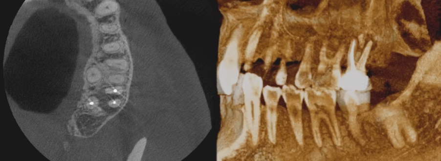
Modern endodontics uses cone beam computed tomography (CBCT), an advanced form of imaging in three dimensions. This technology allows teeth, root canals and surrounding tissues to be examined in far greater detail than with conventional radiographs in two dimensions.
CBCT allows the tooth to be viewed in three dimensions from multiple angles, supporting a more accurate assessment of root canal anatomy, surrounding bone and nearby structures such as sinuses and nerves. This provides valuable information for diagnosis and treatment planning.
Examine the tooth and surrounding tissues in multiple planes.
Gain more information for diagnosis and treatment planning in selected cases.
Scanning is performed when clinically indicated, using an appropriate field of view.
3D imaging can be particularly useful in complex cases, including:
A short looping visualisation illustrates how imaging data can be explored in different views.
Illustrative animation of the CBCT image. This is not an actual recording from scanning software.
Small inflammatory lesions or anatomical features can sometimes be difficult or impossible to identify on conventional radiographs. In selected cases, CBCT can help identify findings more accurately.
A targeted scan and appropriate protocol using a low radiation dose are used to maximise diagnostic value while minimising radiation exposure to the patient.
Explore the uploaded CBCT volume by scrolling through consecutive slices in three anatomical planes. Select a plane and drag the slider to navigate.
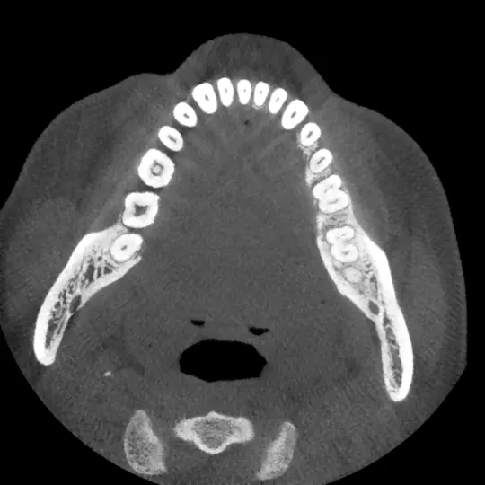
Real CBCT images provided for demonstration. Images are downsampled for the web and must not be used for diagnosis. The original DICOM files are not published.